 |
|
 |
| |
發行人:黃建璋所長 編輯委員:曾雪峰教授 主編:林筱文 發行日期:2022.07.30 |
| |
|
 |
|

本所林清富教授榮獲PARC Appreciation Award,特此恭賀!
帕羅奧多研究中心公司(Palo Alto Research Center, Inc.; PARC),前身為全錄帕羅奧多研究中心(Xerox PARC),是美國的一個科技研發機構,成立於1970年,坐落於加州的帕羅奧多,曾是全錄公司最重要的研究機構。在2002年1月4日起成為獨立子公司(https://www.parc.com/)。
|
|
 |
|
 |
|
| |
|
 |
Speckle noise removal using a two-step weighted robust regression
Professor Jui-che Tsai
Graduate Institute of Photonics and
Optoelectronics, National Taiwan University
臺灣大學光電所 蔡睿哲教授
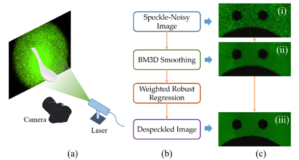
The speckle usually degrades the signal quality for coherent detection or imaging. In this study, under the single-image constraint, we propose a two-step weighted robust regression method for speckle removal, where the first step is to smooth the speckle-noisy image by means of block matching and 3D filtering (BM3D), and the second step is further to apply a weighted robust regression to discriminate between the signal and residual speckle and to recover the edge information. Theoretical demonstration is presented, and experimental results with laser illumination are shown and compared to other competing methods. The experiments show that our method has robustness and prominent improvement of 12~19% in the speckle removal task.
|

|
|
(a) The experimental setup to capture the speckle-noisy images. (b) The flowchart of our two-step weighted regression method. (c) The intermediate images at each step of the flowchart in (b) |
© Elsevier B.V.
H. H. Chen and J. C. Tsai, “Speckle noise removal using a two-step weighted robust regression,”
Optics Communications,
Vol. 452, pp. 510–514, Dec. 2019.
Investigating the contact and high field transport properties of Janus WSSe and MoSSe Materials
Professor Yuh-Renn Wu ’s Laboratory
Graduate Institute of Photonics and
Optoelectronics, National Taiwan University
臺灣大學光電所 吳育任教授
Janus transition metal dichalcogenides TMD with out-of-plane structural asymmetry have attracted increasing attention due to their exceptional potential in electronic and optical applications. Unlike most 2D materials, which has symmetric structure, the Janus TMD has structure asymmetry, which will induce dipole charges in the out-of-plane direction. Hence, in the multi-layer structures, it will behave like piezoelectric material but without surface defects. This enables many device concepts to be implemented with this unique property. For example, the oxide/Janes TMD material interface will induce 2DEG or 2DHG depending on its’ polarity. This will overcome the doping issues in 2D materials, where we don’t need modulation doping to induce n or p channels. A similar comparable device is like AlGaN/GaN HEMT, which is widely used in high-power and high-frequency devices where doping is not needed. The different orientations of multi-layer design also enable multi-function devices. In this study, we systematically investigated the electron-phonon interactions and related transport properties in monolayer Janus MoSSe and WSSe using density-functional formalism. The electrical potential and contact properties were investigated. Our studies show the potential bending at heterojunction interface, which might be used for polar-structure engineering. Furthermore, the electron-phonon scattering rates were obtained using Fermi’s golden rule and extended to the extraction of the effective deformation potential constants for further Monte Carlo treatment. From the results of the Monte Carlo analysis, we found that WSSe provides better performance with higher low-field mobility, while MoSSe shows higher peak velocity at higher fields. In our results, both MoSSe and WSSe seem to be competitive with other previously studied 2D materials. However, with the asymmetric induced piezoelectric effect, it is possible to implement more ideas in device designs. This work provides a systematic perspective on the potential of Janus WSSe and MoSSe for electronic applications.
Partial contents of this report is published in
J. Appl. Phys.
131, 144303 (2022); https://doi.org/10.1063/5.0088593
|
 |
| |
|
|
|
 |
|
| |
|
 |
論文題目:Development of a Nyquist-exceeding subminute gigapixel Nonlinear Optical Mesoscope to assist with rapid histopathology of a human brain tissue(開發超越尼奎斯速率的亞分鐘十億像素介觀顯微鏡以實現人腦組織的快速細胞病理學診斷)
姓名:Bhaskar Jyoti Borah(巴卡地)
指導教授:孫啟光教授
| 摘要 |
|
Intraoperative margin assessment (IMA) is critical in surgical pathology, especially related to vital organs, such as the human brain, which is a crucial part of the central nervous system. Frozen section (FS) is the global standard for IMA, which involves cryosectioning, susceptible to artifacts, consumes up-to 30 minutes per round, and eventually limits the number of IMAs in a critical surgery, which often leads to recurrence of a malignant tumor owing to leftover positive margins. Therefore, a reliable high-speed IMA is always a clear necessity in different levels of surgical pathology applications.
In the paradigm of modern
digital IMA, for a fresh unsectioned tissue, it is often expected to collect ample amounts of information during the IMA itself. The purpose is to digitally secure decent diagnostic reliability so that the specimen does not require to be fixed or deep-frozen and physically preserved for a subsequent re-assessment. Several optical imaging modalities attempted to provide a rapid assessment with optical virtual sectioning of a specimen. Nevertheless, such prior digital IMA-potential studies often might not meet the prevailing state-of-the-art whole-slide-imaging (WSI) standard in the context of either a Nyquist-satisfied centimeter-scale gigapixel-sampled half-a-micron digital resolution, and/or a real-time artifact-free stitching/mosaicking feature, and/or a subminute gigapixel acquisition and digital display ability with an uncompromised resolution across the “specimen to digital display” pixel pathway.
Aside from speed and resolution concerns, the prior FS-alternative IMA approaches often encountered an accuracy/reliability concern. It is noted that pathologists across the globe are trained and adapted to the standard hematoxylin & eosin (H&E)-specific histological features. While adapting to H&E-alternative dyes, especially a source of nuclei contrast other than the gold standard, it is obvious that the results/images produced would not be readily assessed by a pathologist. That eventually needs dedicated deep learning and/or additional interpretation training for a pathologist, while establishing the same degree of accuracy and reliability as the gold standard remains another concern, not to mention repeated organ-specific and/or even hospital-specific training might be required for different surgical pathology applications.
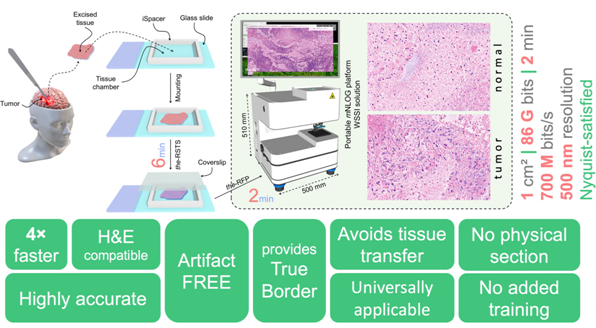
In this research, we introduce an FS-alternative digital IMA-capable technique called Training-Free True-H&E Rapid Fresh
digital-Pathology (the-RFP) that provides 4× faster assessment compared to FS-biopsy, yet holds an excellent reliability comparable to a formalin-fixed paraffin-embedded (FFPE) biopsy. With H&E compatibility,
the-RFP requires no additional interpretation training for a pathologist. Unlike an FS-biopsy,
the-RFP is free from tissue-freezing and physical sectioning, and thus does not encounter typical frozen-section artifacts.
The-RFP is assisted by a mesoscale Nonlinear Optical Gigascope (mNLOG), which provides a truly WSI-competing whole specimen superficial imaging (WSSI) digital IMA-potential solution with an ability of multichannel imaging of a 1 cm2 area in <120 seconds with 86 G bits or 3.6 Gigapixels of data, yielding a sustained effective throughput of >700 M bits/s, preserving a submicron digital resolution with no post-acquisition image processing.
mNLOG platform is streamlined to a rapid artifact-compensated 2D large-field mosaic-stitching (rac2D-LMS) approach yielding a mosaic-stitching ability of >12×12 mm2 area with 130 G bits of data in 60 seconds.
In this study, the-RFP is applied to investigate human brain tumors, being one of the fatal cancers across the globe. Despite unprecedented revolutions in medical sciences and technologies, the 5-year survival for glioblastoma cases only improved from 4% to 7% within the period 1975-1977 to 2009-2015, revealing its extreme seriousness. A fast and reliable IMA is thus critical to effectively resect such a fatal tumor while not much injuring eloquent regions to help circumvent postoperative neurological complications. A non-interventional clinical trial is conducted in collaboration with the Division of Neurosurgery, Department of Surgery, and Department of Pathology of National Taiwan University Hospital to investigate the accuracy and reliability of
the-RFP technique. With no additional interpretation training provided to our collaborating pathologists, blind tests reveal an excellent accuracy of 100% considering a total of 50 normal and tumor-specific human brain specimens, where
the-RFP-based decisions matched the respective FFPE-pathology outcomes.
|
 |
|
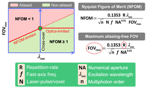
Figure 1. Nyquist figure-of-merit (NFOM) |
|
 |
|
Figure
2. Rapid Fresh
digital-Pathology
(RFP) |
|
|
 |
|
 |
|
| |
|
 |
—
資料提供:影像顯示科技知識平台 (DTKP, Display Technology
Knowledge Platform) —
—
整理:林晃巖教授、吳思潔 —
編織光
量子力學的自旋統計定理(spin-statistics theorem)規定基本粒子必須是玻色子(boson)或費米子(fermion),分別具有整數或半整數值的自旋。在考慮多個可互換粒子的波函數物理學時,玻色子和費米子的表現非常不同。光子作為自旋1粒子,是具有對稱多粒子波函數的玻色子(兩個粒子交換後相移為零),從而產生光子束、玻色子採樣(boson sampling)和玻色-愛因斯坦凝聚(Bose–Einstein condensation)等現象。另一方面,電子是自旋1/2費米子,具有非對稱多粒子波函數(兩個粒子交換後的相移為π),直接導致包立不相容(Pauli exclusion)原理。
然而,這種嚴格的相位條件不再適用於僅限於在兩個空間維度中移動的準粒子[1]。 典型的例子是螺線管磁通量線—就像一縷頭髮—附著在被限制在平面上移動的電子上。根據附加的磁通量,累積相位可以取任意值,這導致Frank Wilczek創造了術語 任意子(anyon)[2]。此外,該相位角取決於準粒子是順時針還是逆時針方向互換,這種區別僅在兩個空間維度上才有意義。
交換兩個粒子的世界線的數學運算在紐結理論(knot theory)中稱為編織。對於幾對粒子的連續應用留下了一個獨特的模式—一個辮子(plait)—它編碼了成對交換的歷史。這類似於頭髮或織物的編織,其中移動和扭曲的順序會產生獨特的圖案(圖1)。數學中的編織是一個純粹的拓撲概念,在理論粒子物理學中有驚人的應用。特別是,它只關心交換或移動的順序,而不考慮它們的精確實現。數學中的紐結理論感興趣的是確定兩個結或辮子是否彼此等價,即它們是否可以連續地從一個轉換到另一個。已經提出了幾個拓撲結不變量,最著名的是瓊斯多項式。然而,計算瓊斯多項式在計算上是困難的,並且有人建議量子計算將提供顯著的計算加速。
|

|
|
圖1、使用光和光波導的非阿貝爾編織原理。一系列緊密間隔的波導以這種方式小心地絞合在一起,以實現與不同位置的不同波導的倏逝耦合(evanescent coupling)。在這種排列中光子的絕熱演化模擬了非阿貝爾任意子的統計數據,因此形成了拓撲量子計算的基石。(我們感謝 V. Neef 對圖的概念設計。) |
最初是在分數量子霍爾效應(quantum Hall effect)[3]的背景下進行研究的,其部分分數統計數據可以串聯,此後任意子已進入量子計算的領域[4],其中它們的拓撲起源本質上可以保護它們免受某些計算錯誤的影響[5]。這也是非阿貝爾任意子(non-Abelian anyon)概念出現的地方。如果透過交換兩個任意子得到的累積相位是一個純量(即一個簡單的數字Θ),那麼這種類型的後續操作是可交換的。這與阿哈羅諾夫-玻姆效應(Aharonov– Bohm effect)沒有什麼不同,在該效應中,帶電粒子的波函數在環繞螺線管磁通線時獲得相位。然而,如果存在一個可以發生演化的能量簡併子空間,則獲得的相位可以是矩陣值的,並且後續操作可能不再可交換。這相當於在曲面,像是地球表面上封閉輪廓上平行傳輸向量,將向量旋轉一個由封閉表面積決定的角度。將向量平行傳輸前後的線性映射可以用旋轉矩陣來表示。正是這些矩陣值運算構成了拓撲量子計算機的基礎。拓撲保護帶來的強健性使得拓撲量子計算的想法非常有吸引力。
在實驗上,已經在最初提出物理學的凝態系統中尋求證明任意子的存在及其編織統計數據[6,7]。現在,Zhang等人在Nature Photonics上發表文章,選擇了用光來追求一條截然不同的路線[8]。基於積體波導結構,在近軸近似下可以模擬離散量子粒子在(2+1)維中的演化,其中光的傳播方向可以作為時間座標,兩橫向作為兩個空間座標。換句話說,將其中一個維度指定為時間,剩下的就是兩個空間維度,不用像固態實現那樣進一步進行維度限制。事實上,這樣的結構代表了粒子的世界線。
使用雷射寫入方法製造光波導可以實現複雜的三維電路幾何形狀,每個波導都遵循自己的特定軌跡。通過明智地選擇相鄰波導之間的間距,以及它們之間的站點間跳躍,他們能夠在其簡併子空間內構建光子的絕熱演化,並決定相關的矩陣值幾何相位 [9,10]。然後透過連接兩個以不同序(order)對相鄰光子成對作用的編織詞來顯示該相位的非阿貝爾性質。如果交換個別運算,兩個串聯操作的結果將是相同的。然而,矩陣值幾何相位的非阿貝爾性質意味著它們的干涉測量產生了複雜的結果。
重要的是,作者還展示了如何利用他們的方案來實現多模非阿貝爾編織。為此,他們使用了一種配置,重複雙模編織的結構作為基本構建。在使用古典雷射以及波長約為810 nm的單光子的概念驗證實驗中,他們展示了五種模態的非阿貝爾編織。由於在這種結構的每個構建區塊中只有一個波導遵循彎曲的軌跡,因此頂部和底部的直波導都是相同的。因此,在未來的應用中,這些可以被預製,並且只有每個中央(即每四個)波導可以基於電路的所需功能來實現。
Zhang等人展示了積體光子波導如何透過使用簡併子空間中光子的絕熱演化來模擬非阿貝爾任意子的統計數據,作為拓撲量子計算的潛在可擴展平台。更有趣的是,這裡沒有涉及到“真正的”準粒子,相反的,光子的演化可以被扭曲成任意子的演化。
儘管這些最新發現只是實現光子拓撲量子計算的第一步,但Zhang和同事[8]提供了一個原理證明演示所有必要的成分—非阿貝爾幾何相位、類任意子統計和級聯運算—可以使用積體光子波導成功實現。現在,積體量子光子學成為拓撲量子計算的可行平台的大門已經敞開。
|
參考資料: |
Stefan Scheel, Alexander Szameit, “A braid for light,”
Nature Photonics volume
16, pages 344-345 (2022)
https://doi.org/10.1038/s41566-022-00993-1
DOI: 10.1038/s41566-022-00993-1
|
|
參考文獻: |
[1] Leinaas, J. M. & Myrheim, J.
Il Nuovo Cimento B 37,
1–23 (1977)
[2] Wilczek, F.
Phys. Rev. Lett. 49,
957 (1982)
[3]
Halperin, B. I. Phys. Rev. Lett.
52, 1583 (1984)
[4] Freedman, M. H., Kitaev, A., Larsen, M. J. & Wang, Z.
Bull. Amer. Math. Soc. 40,
31–38 (2003)
[5] Nayak, C., Simon, S. H., Stern, A., Freedman, M. H. & Das Sarma, S.
Rev. Mod. Phys. 80,
1083 (2008)
[6] Nakamura, J., Liang, S., Gardner, G. C. & Manfra, M. J.
Nat. Phys. 16,
931–936 (2020)
[7] Bartolomei, H. et al. Science
368, 173 (2020)
[8] Zhang, X.-L. et al. Nat. Photon. https://doi.org/10.1038/s41566-
022-00976-2 (2022)
[9] Yang, Y. et al. Science
365, 1021–1025 (2019)
[10] Kremer, M., Teuber, L., Szameit, A. & Scheel,
S. Phys. Rev. Res. 1,
033117 (2019) |
|
|
|
|
|
 |
|
|
|
|
|
|
|
|
|
|
|
 |
|
|
|
 |
|
 |
|