 |
|
 |
| |
发行人:黄建璋所长 编辑委员:曾雪峰教授 主编:林筱文 发行日期:2021.12.30 |
| |
|
 |
|

本所李君浩教授荣膺「2022 SPIE Fellow」,特此恭贺!

本所林恭如教授与阳明交通大学合作研究成果之论文获选发表于「美国光学学会Spotlight
on Optics」,特此恭贺!
论文名称:Ultrafast
2 × 2 green micro-LED array for optical
wireless communication beyond 5 Gbit/s
相关网址:https://www.osapublishing.org/spotlight/summary.cfm?id=460138

本所苏国栋教授荣获「中华民国光电学会2021年度张明文青年光学科技奖」,特此恭贺!
 本所林晃岩教授指导硕士生张乃元同学荣获「The 12th International Conference on 3D Systems and Applications (3DSA 2021) - Best Poster Paper Award」,特此恭贺!

本所林恭如教授指导硕士生翁瑞鸿同学荣获「中华民国光电学会2021年度硕士论文奖」,特此恭贺!
 本所教授指导硕、博士生荣获「
 OPTIC 2021
Student Paper Award」,获奖信息如下,特此恭贺!
|
学生姓名 |
奖项 |
指导教授 |
|
Bhaskar Jyoti Borah(巴卡地)
(博士生) |
OPTIC 2021 Student Paper Award – Oral
论文名称:Sub-Minute Multicolor Giga-Pixel Nonlinear Optical Mesoscope with >30 M/s Effective Pixel Rate |
孙启光 |
|
曾耀賝
(硕士生) |
OPTIC 2021 Student Paper Award – Oral
论文名称:H&E-enhanced nonlinear optical microscopy to rapidly assess glioma border in human brain |
孙启光 |
|
钱子群
(硕士生) |
OPTIC 2021 Student Paper Award – Oral
论文名称:One-bit Transmission Analyzation of asymmetric-arm Silicon Photonic Mach–Zehnder Modulator |
林恭如 |
|
张雨倢
(硕士生) |
OPTIC 2021 Student Paper Award – Oral
论文名称:Analysis of TM polarization ratio in UVC-LEDs with 3D k•p method by considering random alloy fluctuation |
吴育任 |
|
薛陆清
(硕士生) |
OPTIC 2021 Student Paper Award – Oral
论文名称:Development of Integrated Optoelectrical Amplifier and High Resolution Smart Thermal Sensor with Light-Emitting Transistors |
吴肇欣 |
|
蔡侑男
(博士生) |
OPTIC 2021 Student Paper Award – Poster
论文名称:Cellular imaging with a customized spectral-domain OCT system and an in-house developed cell incubator |
李翔杰 |
|
张浩升
(硕士生) |
OPTIC 2021 Student Paper Award – Poster
论文名称:Design and Realization of Wide Field-of-view 3D MEMS LiDAR |
苏国栋 |
|
|
 |
|
 |
|
| |
|
|
|
|
|
 |
News on Optoelectronics
2021 Queen Elizabeth Prize for Engineering
GIPO is delighted to share the news of 2021 Queen Elizabeth Prize. Several of GIPO’s faculty members, including Prof. Jian-Jang Huang, Prof. Chao-Hsin Wu and Prof. Yuh-Renn Wu, have long term collaborations and interactions with the recipients, Prof. Nick Holonyak Jr, Prof. Russell Dupuis, Dr. M. George Craford and Prof. Shuji Nakamura.
LED lighting development wins 2021 Queen Elizabeth Prize for Engineering.
Isamu Akasaki, Shuji Nakamura, Nick Holonyak Jr, M. George Craford and Russell Dupuis have been awarded the world's most prestigious engineering accolade.
First awarded in 2013 in the name of Her Majesty The Queen, the QEPrize exists to celebrate ground-breaking innovation in engineering. The 2021 winners were announced on February 2 by Lord Browne of Madingley, Chairman of the Queen Elizabeth Prize for Engineering Foundation.
Solid-state lighting technology has changed how we illuminate our world. It can be found everywhere from sports stadiums, parking garages, inside and outside commercial buildings, homes, digital displays and computer screens and cell phones to hand-held laser pointers, automobile headlights and traffic lights. Today’s high-performance LEDs are used in efficient solid-state lighting products across the world and are contributing to the sustainable development of world economies by reducing energy consumption.
Visible LEDs are now a global industry predicted to be worth over $108 billion by 2025 through low-cost, high-efficiency lighting. They are playing a crucial role in reducing carbon dioxide emissions, consuming significantly less energy and producing 90% less heat than incandescent lighting, and their large-scale use reduces the energy demand required to cool buildings. For this, they are often referred to as the ‘green revolution’ within lighting.
Link
News referred from:
https://qeprize.org/winners
|
 |
|
|
|
|
|
|
|
|
|
 |
|
| |
|
 |
|
林建中教授于2002年取得美国史丹佛大学电机工程博士学位,指导教授为Professor James Harris。其博士论文主要在半导体光电组件与微机电结构(MEMS)的单石化整合(monolithic integration)上。毕业之后投入美国相关的新创公司工作,包括E2O Communication Inc.以及Santur Corp.两家新创公司。期间由一般的研发工程师逐步升至相关的经理职位。工作范围包括了长波长VCSEL激光以及波长可调变之DFB激光数组。这些组件,在当时不但属于新颖技术,也是业界极为需要商品化的关键光通讯组件。2009年林博士返国加入国立交通大学光电学院光电系统研究所,重新返回学术单位,进行相关的光电组件研究。之后在2021年8月加入国立台湾大学电机系及光电工程学研究所任教。
林教授的研究领域着重于新颖光电组件的制作与设计。包括高效率量子点发光与光侦测组件、微小化发光二极管应用于微显示屏幕、多重发光之颜色转换层应用于光电组件等等方向。涵盖了相关的材料研究以及组件的制程与封装。同时也对于如何延长新颖材料的使用寿命,进行广泛而深入的探讨。
林教授相关的研究成果多发表于国际顶尖会议与学术期刊,如:CLEO、IEEE IPC、IEEE PVSC、Advanced Functional Materials、IEEE JSTQE、Optics Express等,历年来已发表超过230篇的相关论文。林教授是科技部优秀年轻学者研究计划得主,同时也于2020年荣获美国光学学会会士的荣誉。
林教授目前参与的计划以前瞻显示计划、硅光子同调通讯计划为主,并且主持科技部个人型研发计划,着力于利用量子点强化传统半导体太阳能电池组件的功能。
在教学研究之余,林教授的兴趣是看老电影。
林教授认为,光电对于学生来说,是一个介于基础科学(如物理、化学、生物)与工程(如半导体、通讯)之间的学科。也因为如此,对于跨学门的知识与训练特别重要。尤其光子与电子的性质迥异,许多的新领域尚待探索。林教授相信在后摩尔时代里,光电的重要性将会与日俱增,非常值得有兴趣的同学投入。林教授也鼓励同学,学习的秘方无他,就是持之以恒,努力不懈。
|
| |
|
 |
|
 |
|
| |
|
 |
Imaging melanin quantitatively with a high 3D spatial resolution in human skin
Professor Chi-Kuang Sun
Graduate Institute of Photonics and
Optoelectronics, National Taiwan University
台湾大学光电所 孙启光教授
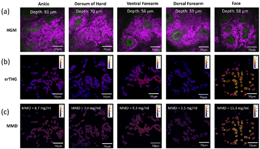
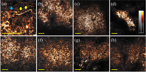
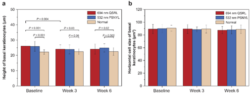
The capability to image the 3D distribution of melanin in human skin in vivo with absolute quantities and microscopic details will not only enable noninvasive histopathological diagnosis of melanin-related cutaneous disorders, but also make long-term treatment assessment possible. Recently, we developed the label-free third-harmonic-generation (THG) enhancement-ratio microscopy and demonstrated clinical in vivo imaging of the melanin distribution in human skin with absolute quantities on mass density and with microscopic details [1]. As the dominant absorber in skin, melanin provides the strongest THG nonlinearity in human skin due to resonance enhancement. We show that the THG-enhancement-ratio (erTHG) parameter can be calibrated in vivo and can indicate the melanin mass density (Fig 1). With an unprecedented clinical imaging resolution, we further applied the erTHG-microscopy’s unique capability for long-term treatment assessment in solar lentigines and melasma, and direct clinical observation of melanin’s micro-distribution, cellular morphological changes and other in vivo histological morphologic parameters. We observed the melanocytes remain hyperactivation after treatment in melasma patients after treatments (Fig 2), and the height of basal keratinocytes remain abnormal in SLs patients after laser treatment (Fig 3). Our study indicates that these parameters can serve as clinical indices to evaluate the histopathological characteristics of pigmentary disorder patients and the effectiveness of treatments. All the clinical research outcomes have been published in
IEEE journal of selected topics in quantum electronics and
Biomedical Optical Express [1-3].
|
 |
|
Fig. 1. Examples of melanin mass density (MMD) and erTHG images. (a). The slide-free in vivo HGM images en-face optically sectioned underneath the skins of dorsal forearm, ventral forearm, ankle, dorsum of hand, and face of different volunteers. (b). The corresponding erTHG images of the segmented cell cytoplasm. (c). The corresponding MMD distribution images inside the cytoplasm of basal cells. Epi-SHG and epi-THG are represented by green and magenta pseudo-colors. erTHG, MMD distribution images are color coded. |
|
 |
|
Fig.2 Representative in vivo THG human skin images with different melanocyte dendricity scores (MDS) in melsam patients. (a) A typical melanocyte in the THG human skin image with a cell body (blue arrow) and an obvious long dendrite (yellow arrow). (b)(c)(d) Examples of THG human skin images with the lowest MDS equivalent to 0. (e)(f)(g)(h) Examples of THG human skin images with the highest MDS equivalent to 3. Scale bar = 50 µm |
|
 |
|
Fig.3 Morphological parameters relevant to basal keratinocytes in SLs patients. The (a) HBK and (b) HCS of basal keratinocytes were analyzed from the en face in vivo HGM images at baseline, week 3, and week 6. Statistical differences were tested using the paired t-test. PSNYL: picosecond Nd:YAG laser; QSRL: Q-switched ruby laser; HBK: height of basal keratinocytes; HCS: horizontal cell size; HGM: harmonic generation microscopy. |
Reference:
[1] Sun, C. K., Wu, P. J., Chen, S. T., Su, Y. H., Wei, M. L., Wang, C. Y., ... & Liao, Y. H. (2020). Slide-free clinical imaging of melanin with absolute quantities using label-free third-harmonic-generation enhancement-ratio microscopy. Biomedical Optics Express, 11(6), 3009-3024.
[2] Wei, M. L., Liao, Y. H., Weng, W. H., Shih, Y. T., Sheen, Y. S., & Sun, C. K. (2021). A Study on Applying Slide-Free Label-Free Harmonic Generation Microscopy For Noninvasive Assessment of Melasma Treatments With Histopathological Parameters. IEEE Journal of Selected Topics in Quantum Electronics, 27(4), 1-10.
[3] Wu, P. J., Chen, S. T., Liao, Y. H., & Sun, C. K. (2021). In vivo harmonic generation microscopy for monitoring the height of basal keratinocytes in solar lentigines after laser depigmentation treatment. Biomedical Optics Express, 12(10), 6129-6142.
Dual-depth Augmented Reality Head-up Display with Holographic Imaging
Professor Hoang-Yan Lin
Graduate Institute of Photonics and
Optoelectronics, National Taiwan University
台湾大学光电所 林晃岩教授
Head up displays (HUDs) show useful information on a combiner or windshield of a vehicle so that drivers do not need to look down on their dashboards and can focus on the road. As HUDs are proved to increase drivers’ safety and driving experience, HUDs have become more popular over the past decade. Conventionally, head up displays use liquid crystal displays (LCDs) as their picture generation units (PGUs). Most commercial HUDs are LCD-based and project a single image on its combiner. However, as the concept of augmented reality and virtual reality have become well-known and popular, with the upgraded computational speed and performance, augmented reality head up displays (ARHUDs) have become the future of HUDs.
Compared to traditional HUDs, ARHUDs can be designed to provide at least two virtual images with different depths, with at least one virtual image interacting with real world, so that drivers can have immediate reaction without visual conflicts. A straightforward method to realize this concept is to use two or more LCDs as PGUs and corresponding imaging optics to produce several virtual images at different depths. However, this method requires much more cost and volume, which makes it unsuitable for the scenario of HUDs.
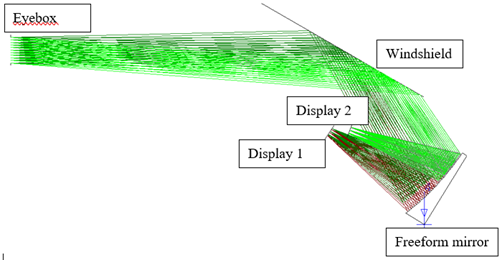
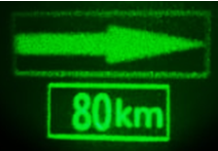
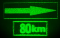
As shown in Fig. 1, we develop a dual-focal-plane ARHUD based on holographic imaging, which allows dynamic depths of reconstructed images by varying the modulation of a spatial light modulator (SLM), e.g. liquid crystal on silicon (LCOS). With holographic imaging system in replace of LCDs as PGUs, A single freeform mirror is designed to further magnify the projection sizes and depths, which fits the simultaneous projection of two separated images, reconstructed at two different depths by holographic projection system, producing a far image (15° x 3° at 7 m) and a near image (6° x 2° at 2.5 m), as shown in Fig. 2, in an eyebox of 100 mm by 60 mm. An algorithm on distortion correction based on Code V analysis also shows effective result of correcting the complex and non-radial distortion of freeform off-axis optical system.
|
 |
|
(a) Illustration of the specifications. |
|
 |
|
(b) Layout or magnification of highlighted portion in (a). |
|
Fig. 1. Specification and layout of the HUD. |
|
 |
|
(a) (b) |
|
Fig. 2. Holographic reconstruction of two images with two different depths (a) focused at the near icon, 80 km; (b) focused at the far icon, arrow. |
Reference:
I-Chen Cho, You-Ming Hu, Chih-Hao Chuang, Chi-Wen Lin, Kuan-Hsu Fan-Chiang, and Hoang-Yan Lin, Dual-depth Augmented Reality Head-up Display with Holographic Imaging, 3DSA 2021.
|
 |
| |
|
|
|
 |
|
| |
|
 |
论文题目:可挠性氧化物半导体放大电路及其于生物讯号感测之应用
姓名:许书铭
指导教授:陈奕君教授
| 摘要 |
|
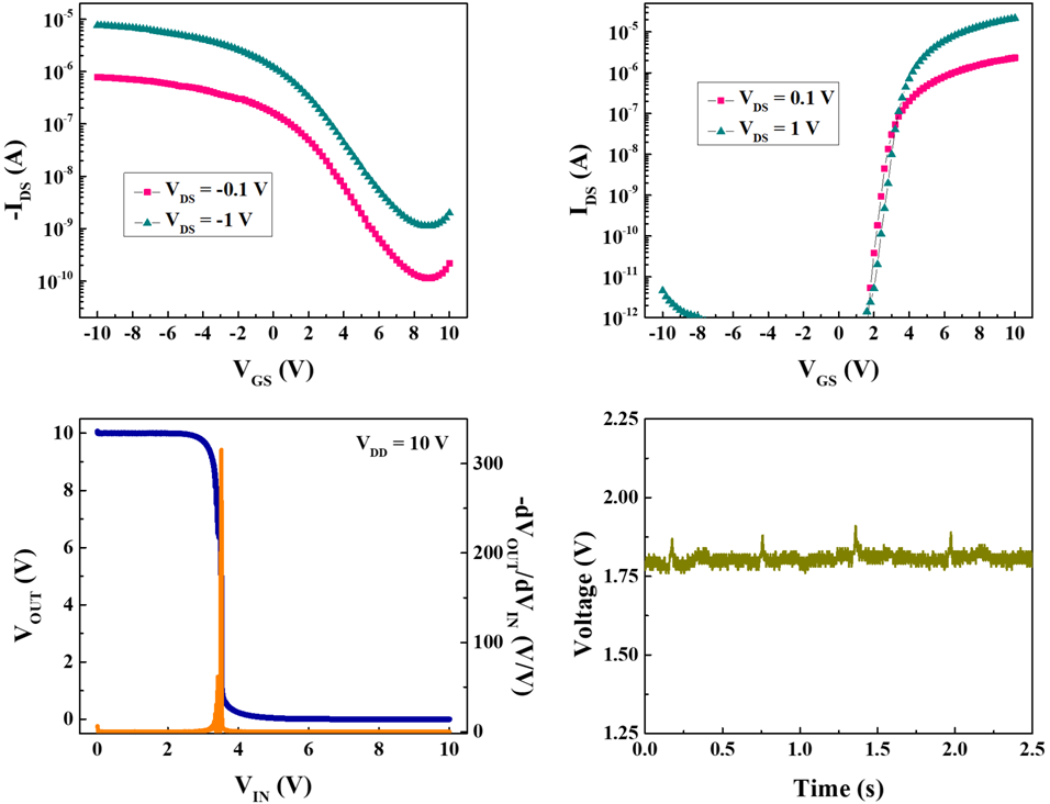
可挠性电子(flexible electronics)技术因具有轻、薄,以及可弯曲等特性,是实现肌肤电子(skin electronics)产品的关键技术。目前大多数可挠性电子组件皆制作于塑料基板上,考虑塑料所能承受之制程温度,能在相对低温下制造的氧化物半导体(oxide semiconductor),是适合开发可挠性电子组件的半导体材料。本研究首先于聚酰亚胺(polyimide)基板上开发P型氧化亚锡(tin monoxide)和N型氧化铟镓锌(indium gallium zinc oxide)薄膜晶体管(thin-film transistor),并结合原子层沉积系统之覆盖效应(atomic-layer-deposition capping effect),优化晶体管之电性表现。接着,整合氧化亚锡和氧化铟镓锌薄膜晶体管开发CMOS反相器(complementary metal-oxide-semiconductor inverter),透过调整CMOS的结构和制程之优化,成功使CMOS反相器的静态电压增益于供应电压10 V时能超越300 V/V,同时组件展现优异的弯曲可靠性,不论是在受到张应变还是压应变的情况下,即便弯曲半径小至2.5 mm,整体电性皆无明显之变化。透过此可挠性CMOS技术,我们成功制作了CMOS差分放大器,其差模增益值、共模增益值,以及共模互斥比分别约为51 V/V、8.5 V/V和15.6 dB。最后将市售之心电量测电极贴附于胸口,并连接至可挠性CMOS差分放大器,成功读取到了心电讯号,此结果证明了所开发之基于氧化物半导体的可挠性电子技术,在具有生物讯号监测功能之肌肤电子产品上的应用潜力。
|
 |
|
图一、可挠性CMOS反相器的(a)示意图及(b)照片。[1] |
|
 |
|
图二、可挠性(a)P型氧化亚锡和(b)N型氧化铟镓锌之转换特性曲线。(c)可挠性CMOS反相器之电压转换特性。(d)透过可挠性CMOS差分放大器所得到之心电讯号。 |
Reference:
[1] S.-M. Hsu, D.-Y. Su, F.-Y. Tsai, J.-Z. Chen, and I.-C. Cheng, “Flexible complementary oxide thin-film transistor-based inverter with high gain,”
IEEE Trans. Electron Devices, vol. 68, no. 3, pp. 1070-1074, Mar. 2021, doi: 10.1109/TED.2021.3052443. |
|
 |
|
 |
|
| |
|
 |
— 资料提供:影像显示科技知识平台 (DTKP, Display Technology
Knowledge Platform) —
—
整理:林晃岩教授、吴思洁 —
太赫兹波之相位控制移往芯片上
太赫兹(Terahertz , THz)辐射,通常称为T波,在微波和红外波段之间的电磁频谱中占据独特的位置。随着频率范围从0.1到10 THz,T波在无线电天文学(radio astronomy)和太空科学(space science)中一直是很重要的。然而,随着检测器发展的进步,最重要的是太赫兹辐射源在光谱学、材料特性检测、遥测和诊断、成像和断层扫描[1]等应用上,促进了在研究实验室和工业中对于使用T波的更广泛兴趣;特别是对于生命科学和医学方面,T波有很大的希望,因为许多生物之重要大分子的集体振动是发生在太赫兹频率的范围[2]。此外,太赫兹频率范围被考虑作为下一代高速数据运行的短距离无线系统,并可以与光纤通讯系统衔接 [3]。
然而,因为缺乏用于动态控制和调制T波的高效率和紧凑组件,THz技术的发展及其采用一直受到阻碍,这一功能仍然是一个关键挑战[3]。困难主要是因为T波与天然非金属材料的交互作用很弱,除了最值得注意的掺杂半导体和极性液体(像是水),而它们仅仅是被吸收。
目前采用的主流解决方案之一是提升T波与物质间的相互作用,特别是透过利用人工工程材料,所谓的超颖材料(metamaterial)[4]的电磁共振。这种方法已经能够演示基于主动超颖材料的组件,例如用于T波在自由空间中作为准直或聚焦光束传播的开关和调制器,(例如参考文献[5]-[7])。 然而,迄今为止,这类设计还不能轻易缩小到在太赫兹通信中的芯片级生物医学传感器和数据处理系统[3],其所设想之积体化太赫兹组件的尺寸,更别提新兴太赫兹电子产品所需的微型化程度了。
现在Nature Photonics中报导了,Hongxin Zeng及其团队设计并透过实验验证了太赫兹电路的一个重要组件—芯片上的一种紧凑型高精度电子移相器(phase shifter),它能够对T波进行可变量位编码相位调制[9]。所演示的相位编码芯片的其它显著特点是它实际上不会影响T波的振幅。此外,它展现出低传输损耗(不同于以前利用共振相互作用的解决方案)。
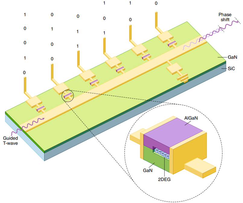
相位编码芯片(如图一)是基于传统微带波导,其中一侧透过微制造的平面电容器连接到一系列沿波导等距分布的六个相同的短金属贴片。在大约 0.3 THz 的频率下,由此产生的微结构(在微波工程中称为短截线(stub))作为微型电磁谐振器,每个谐振器都会局部改变波导的阻抗,因此影响引导T波的传播。它们代表了太赫兹频率范围,等效于电子学中常见的LC谐振电路,而LC谐振电路在如此高的频率下是不可能实现的。由于短截线的谐振是来自驻波的激发,因此可以透过改变短截线的有效电长度来改变谐振的频谱位置(以及它在特定频率造成的振幅变化)。作者透过使短截线中的微电容器可藉由各自的短截线单独提供的控制信号,进行数字切换来实现这一点。结果,他们能够将任何给定短截线的共振进行蓝移高达20%。
|
 |
|
图一、太赫兹移相芯片的原理图,及其如何将数字编码相位调制传递给沿中心波导传播的T 波。相移是通过沿波导上侧放置的短截线引入的,并充当由具有二维电子气(2D electron gas, 2DEG)的微电容器控制的微小太赫兹谐振器。 |
AlGaN/GaN的异质接面(跨越他们的间隙)整合至微电容器的结构是实现大的频谱偏移的关键。已知一个AlGaN/GaN的异质接面会自行产生二维电子气(2D electron gas, 2DEG),两种材料之间的界面处捕获的单层密集电子[10]。2DEG中的电子可以沿着表面自由移动,有效地形成了一个导电片,它几乎完全连接了从一块平板延伸到另一块平板的制造微电容器中的间隙(见图一)。透过间隙施加偏压,可以使2DEG更进一步远离平板之一,从而降低结构中的电容。作者发现将偏压从0 V(状态“1”)切换到 7 V(状态“0”)后电容降低了十倍以上。
为避免T波的振幅变化,会伴随着电子控制的谐振短截线引起的相位变化,Hongxin Zeng及其团队选择在频率范围为0.26–0.27太赫兹、非谐振的状态运行他们的芯片。
这种技巧使他们能够将芯片的整体传输维持在相对较高的水平(–6到–8 dB,取决于频率),波动仅限于0.5 dB。尽管在此频率范围内透过任何单个短截线获得的相移自然小于谐振时(低于10度),但它会随着导波从一个短截线传播到另一个短截线而累积,并可能高达55度。重要的是,短截线单独施加的相移不会仅仅由于后者之间的相互作用而累加,这使得许多(其它等效的)开关组合显得独一无二。例如,输入代码 100001、010010和001100给芯片将产生不同的相移。为了完全解除简并态(degeneracy),Hongxin Zeng及其团队使用了另一个技巧—将控制信号的接地电极作为额外的“无源”短截线引入,他们将其放置在微带的另一侧,更靠近其末端(图一)。微带产生的结构不平衡能确保所有控制短截线,即使单独切换,对引导 T波也有独特的影响—即代码 100000、010000、001000、000100、000010、000001 将编程不同的相移。上述所有代码1的状态将对应至不同的输出。
实施的方法效果很好,使作者能够以数字方式控制 T 波的相位,并且将它以2-5度(取决于操作频率)的小恒定增量偏移,平均误差仅有0.36度。假设一个基于2DEG 的微电容器的开关时间非常短为100 fs,相位编码芯片的调制速率可能高达 3 Gbps,正如最近的一项研究表明的那样[11]。这种高速结合小尺寸芯片(主动区面积为2.3×0.3 mm2)及其“数字化就绪”,使得演示的移相器作为未来积体化太赫兹系统的主动组件特别地有吸引力。
然而,它的实际使用可能仍会被一些留着的问题延迟。特别像是移相器的操作本质上是窄带的,因为它(虽然不是直接)依赖谐振,因此最小以及总可达到的相移两者都取决于工作频率。预计芯片的传输损耗与活性短截线的数量成比例增加,这会限制所提出的相位调制方案的扩展性,实际上,目前展示最大相移30-55度是可能的最佳输出。最后,目前已经针对太赫兹频带的低频边缘演示了芯片上主动式的相位控制,同时对于在1太赫兹及以上的频率振荡的T波,是否能实现相同的工作原理还有待观察。
|
参考资料: |
Vassili Fedotov, “Phase control of terahertz waves moves on chip,”
Nature Photonics 15,
715-716 (2021)
https://doi.org/10.1038/s41566-021-00887-8
DOI: 10.1038/s41566-021-00887-8
|
|
参考文献: |
[1] Ferguson, B. & Zhang, X. C.Nat. Mater.
1, 26–33 (2002).
[2] Liu, W. et al.
Front. Lab. Med. 2, 127–133 (2018).
[3] Tonouchi, M.
Nat. Photon. 1, 97–105 (2007).
[4]
Fedotov, V. in Springer Handbook of Electronic and Photonic Materials 2nd edn (eds Kasap, S. & Capper, P.) Ch. 56, 1351–1375 (Springer, 2017).
[5]
Chen, H. T. et al. Nature
444, 597–600 (2006).
[6]
Savo, S., Shrekenhamer, D. & Padilla, W. J.
Adv. Opt. Mater. 2, 275–279 (2014).
[7] Pitchappa, P. et al. Adv. Mater. 31, 1808157 (2019).
[8] Sengupta, K., Nagatsuma, T. & Mittleman, D. M. Nat. Electron.
1, 622–635 (2018).
[9] Zeng, H. et al. Nat. Photon. https://doi.org/10.1038/s41566-021- 00851-6 (2021).
[10] Ibbetson, J. P. et al. Appl. Phys. Lett. 77, 250–252 (2000).
[11] Zhao, Y. et al. Nano Lett.
19, 7588–7597 (2019).
|
|
|
|
|
|
 |
|
|
|
|
|
|
|
|
|
|
|
 |
|
|
|
 |
|
 |
|